Electron Microscopy
The electron microscopy (EM) laboratory is an integral component of the Department of Pathology and Genomic Medicine at the Houston Methodist Hospital. The EM laboratory is supported by the most up-to-date equipment and facilities that provide all diagnostic services related to both transmission and scanning electron microscopy. The EM laboratory processes more than 800 cases each year. Kidney biopsies constitute the majority of the samples (95%) with the remainder consisting of other types of tissue biopsies including heart, muscle, nerve, and cornea. EM services are also available for research purposes. The average turn around time for clinical EM samples is one day.
Equipment:
JEOL 1400 electron microscope equipped with AMT digital camera
Ultramicrotomes: Leica EM UC7 and RMC PT-XL
PELCO BioWave Pro microwave tissue processor
Lynx2 Automated Tissue Processor
Services:
Complete EM (processing, sectioning, TEM imaging)
Bloc-and-hold EM samples (processing only)
“As-needed” utilization of the electron microscope with technical help by an EM technical specialist
Contact:
EM laboratory: 346.238.7227
electronmicroscopyservices@houstonmethodist.org
Locations:
EM processing lab: WT2-2021, 6551 Bertner Ave, Houston, TX, 77030
Director: Luan Truong, MD
EM Technologists: Mary Hsiao, Huie Wang and Clair Haueter, CEMT
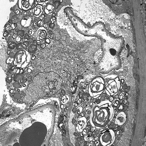
Zebra bodies in Fabry disease