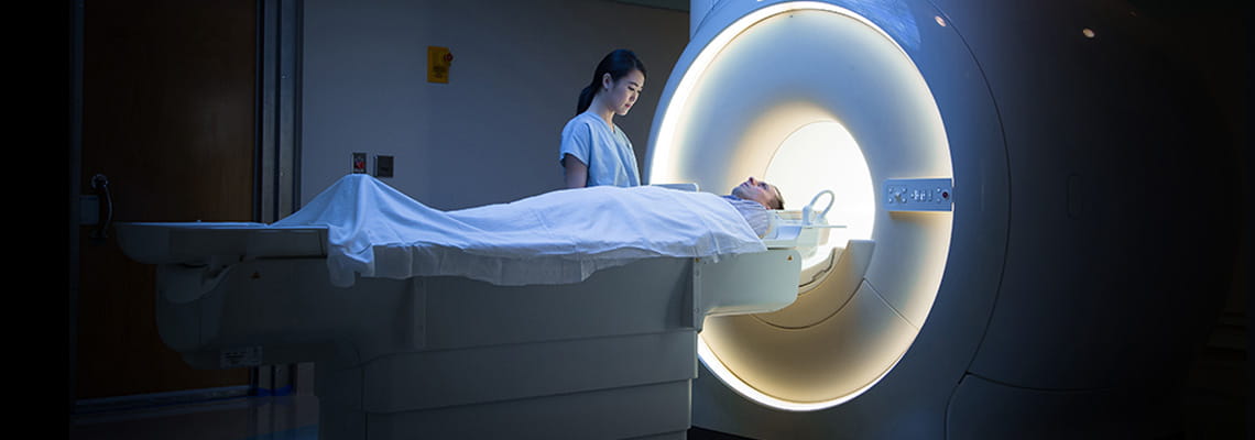
Houston Methodist offers patients advanced imaging and radiology services and technologies. Our board-certified radiologists have access to a range of diagnostic imaging and radiology services, including:
Computed tomography (CT) — moves an X-ray around a patient to produce multiple detailed images from different angles. CT provides greater detail of:
Diagnose brain, spinal cord, bone and joint conditions and injuries
Evaluate infections
Nuclear medicine — uses small amounts of radioactive materials texamine organ function and structure. Positron emission tomography (PET) is a type of nuclear medicine that uses radioactive materials, a special camera and computer tevaluate organ and tissue functions. Tests include:
Bone scan — diagnoses bone diseases, infections or injuries
Brain scan — evaluates brain disorders such as Alzheimer’s disease, seizures, tumors and strokes
Hepatobiliary scan — identifies gallbladder disorders and bile duct obstructions
Lung ventilation and perfusion scan (VQ scan) — measures air and blood flow in and out of the lungs
Lymphoscintigraphy — evaluates the lymphatic system and identifies lymph nodes for removal
Octreoscan — whole-body scans that identify rare neuroendocrine tumors
Renal scan — evaluates Kidney blood flow and function
Parathyroid scan — identifies abnormal parathyroid glands that cause hyperparathyroidism
I-131 whole body scan —detects thyroid cancer spread
Ultrasound — uses sound waves tproduce images of soft tissues inside the body. Types include:
Abdominal ultrasound — detects gallstones or tumors
X-ray — uses a small dose of radiation tproduce images of body structures. Types include:
Barium X-ray — diagnoses conditions such as tumors, polyps, hernias, strictures, ulcers and other gastrointestinal inflammatory conditions in the GI tract
Fluoroscopy — studies movement of internal body parts and systems such as blood flow through a blood vessel or food passing through the stomach and intestines