

Focused microlearning can help clinicians strengthen echocardiographic assessment of the interatrial septum and improve detection of atrial septal defects.
Article Highlights
- Houston Methodist has launched a microlearning echocardiography video series focused on adult congenital heart disease, beginning with interatrial septum assessment and atrial septal defect (ASD) evaluation.
- The inaugural tutorial demonstrates transthoracic echocardiographic techniques for identifying ostium secundum ASDs, a common congenital defect that may be underdiagnosed by TTE.
- Instruction covers optimal imaging windows, including parasternal, apical four-chamber and subcostal views, along with sweep techniques to improve ASD detection.
- Case-based examples highlight the assessment of shunt direction, Doppler sampling and frame-rate optimization across varying ASD sizes and pulmonary pressure conditions.
- The series provides concise, practical echocardiography education for cardiologists, sonographers and other clinicians involved in congenital heart disease care.
The Houston Methodist DeBakey Heart & Vascular Center has debuted a new microlearning video series aimed at helping echocardiographers sharpen specific skills in adult congenital heart disease, beginning with a focused tutorial on interatrial septum assessment and atrial septal defects (ASDs).
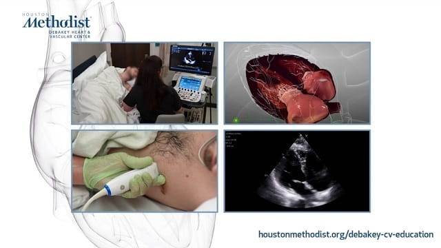
The inaugural 10-minute video features Houston Methodist echocardiography educator Alicia Rangosch demonstrating transthoracic echocardiographic (TTE) techniques for evaluating the interatrial septum. The emphasis is on detecting ostium secundum ASDs, one of the most common but often underdiagnosed congenital heart defects by TTE.
"These brief, high-yield tutorials are designed for cardiologists, sonographers and general practitioners who want to build core echocardiographic skills," said Dr. Miguel Quinones, one of the faculty contributors to the video series.
ASDs occur when there is an abnormal opening in the wall between the heart's two upper chambers, allowing blood to flow between the atria. Ostium secundum ASD, the most frequent subtype, stems from an incomplete closure of the septum primum over the fossa ovalis. While small ASDs may remain asymptomatic, larger or undiagnosed defects can lead to serious complications such as right heart enlargement, arrhythmias, strokes or pulmonary hypertension.
The video walks viewers through preferred imaging windows and techniques — including left and right parasternals long and short axis, apical four-chamber, subcostal views and the use of sweeps — with both simulator-based 3D models and real-time 2D echo footage. Case examples illustrate small and large ASDs under varying pulmonary pressures, highlighting the importance of shunt direction, Doppler sampling and frame-rate optimization.
This is the first installment in a larger digital learning library, reinforcing Houston Methodist's commitment to advancing cardiovascular education through practical, accessible tools. Additional resources are available through the Methodist DeBakey Cardiovascular Journal.