

A surgical overview of PAPVR repair, highlighting key anatomy, hemodynamics and the decision-making behind double patch and Warden techniques.
Video Highlights
PAPVR most commonly involves right-sided pulmonary veins draining into the SVC, often with a sinus venosus ASD. Surgical repair is typically considered when the left-to-right shunt (QP/QS) is significant, reinforcing the importance of accurate anatomical and hemodynamic assessment.
Double patch technique uses an existing (or surgically created) ASD to channel pulmonary venous return into the left atrium while enlarging the SVC to prevent obstruction. Understanding pressure gradients and the expected patch bulge toward the right atrium is critical for planning.
By transecting the SVC and anastomosing it to the right atrial appendage, the Warden technique separates systemic and pulmonary venous pathways. Proper preparation of the appendage is essential to ensure unobstructed venous return.
Cannulation strategy and exposure are central to operative success.
Adequate control of the SVC and venous drainage during cardiopulmonary bypass — often via cannulation at the innominate vein or SVC junction — is key to maintaining operative visualization and flexibility.
Careful attention to the atrial node location, precise ASD creation at the fossa ovalis, and ongoing evaluation of tricuspid regurgitation and pulmonary pressures are critical safeguards during repair.
Partial anomalous pulmonary venous return (PAPVR) is a relatively common form of congenital heart disease with important implications for surgical planning. In this whiteboard review, Dr. Andrea Quarti, surgical director of the Adult Congenital Heart Disease (ACHD) program at Houston Methodist, outlines the core anatomical patterns and hemodynamic drivers that guide intervention. He also compares two primary repair strategies — the double patch and Warden techniques — emphasizing technical considerations and intraoperative decision points. The session concludes with key surgical reminders to optimize outcomes and avoid complications.
Article Transcript
Welcome to the whiteboard lesson on congenital heart disease. My name is Andrea Quarti. I'm the surgical director of the ACHD program at Houston Methodist. The lesson today will focus on a very common disease that is called partial anomalous pulmonary venous return.
We will focus on this disease using two main drawings.
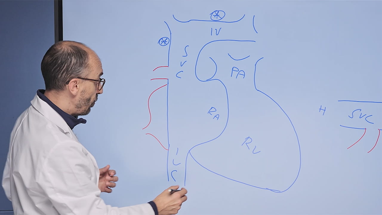
The patient standing. So, innominate vein, SVC, IVC, right atrium, right ventricle, and pulmonary artery.
And with the patient lying on a bed. Here we have the head, the feet, SVC, right atrium, IVC, and left atrium.
In the PAPVR, usually one or more pulmonary veins from the right side are draining into the SVC. There could be an ASD that is called sinus venosus ASD, or there could be an intact interatrial septum.
Supposing the patient needs a surgical repair, in this case the QPQS, so the shunt across the pulmonary vein and, in case the ASD is over 1.5. Supposing we have to do a surgical correction, usually we can use two surgical techniques. One is called the double patch, and one is called Warden.
Double patch technique. The access to the heart can be from the front using a full sternotomy or using a right mini-thoracotomy. Usually I prefer the third intercostal space since we need to have a full control of the SVC. To go on cardiopulmonary bypass, we need a full venous drainage, and this can be accomplished putting a cannula in the SVC at the junction with the innominate vein or into the innominate vein. This is a very flexible and useful idea to have a CPB in these patients.
The abdomen and the legs will be drained using a cannula into the IVC. If there is an ASD, you can use the existing ASD to do the correction. But if there's no ASD, you need to create an ASD and be aware the only place where you can create an ASD is the fossa ovalis. But today we are lucky and we have an ASD in this patient.
It's quite intuitive. The double patch allows us to connect the pulmonary vein to the left atrium using the existent ASD. In this way, the flow will go into the left atrium using the existent ASD.
Let's have a look on this side. This is the ASD and we can put a patch this way. So the flow will go into the left atrium. Obviously, you should know that the pressure into the left atrium is higher compared to the pressure in the right atrium. So this patch will bulge into the SVC. That's why it's called double patch technique.
To avoid an SVC syndrome, it's very important to make an incision in the SVC and to enlarge the SVC. So the SVC can be opened longitudinally. And a longitudinal patch can be placed. In this way, there will be enough space for the venous flow from the pulmonary vein through the left atrium and enough space for the venous return from the head and the arms into the right atrium.
But let's move to the Warden technique. To understand better the Warden technique, I will use this type of drawing. In the Warden technique, that is really amazing, it is possible to transect the SVC at this level and to connect the pulmonary vein and the ASD putting a patch this way.
The SVC will be transected. This way, it is possible to put a suture here and connect the SVC to the appendage. It is possible to open the appendage. Beware to remove all the muscle from inside the appendage to create an unobstructed pathway. And the SVC will be connected to the appendage with the suture.
At the end of the procedure, the SVC will drain into the right atrium and the pulmonary veins will drain into the left atrium.
Now you have the basics to correct the PAPVR, but I want to give you some more details.
First, beware, here is located the atrial node. Whenever you are opening the SVC, be sure that you are very low at this level.
Second, if you need to do an atrial septostomy, be aware that the only place where the left atrium is really touching the right atrium is at the level of the fossa ovalis.
Third, you need always to control the tricuspid valve level of regurgitation and the pulmonary pressure.
Fourth, remind that the patch will bulge toward the right side.
And finally, be safe for your patients. Thank you.