
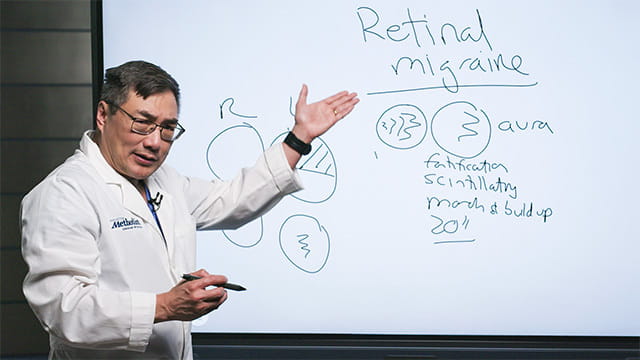

Retinal migraine should be treated as a diagnosis of exclusion, with visual field testing and vascular workups used to rule out other causes.
Article Highlights
- Retinal migraine is defined by transient monocular visual symptoms, distinguishing it from typical binocular migraine aura.
- Diagnostic criteria require fully reversible, short-lived visual loss followed by a migraine headache, with no residual field deficit.
- Retinal migraine should be treated as a diagnosis of exclusion, requiring evaluation for intracranial pathology when visual field patterns are atypical.
- Visual field defects respecting the vertical meridian (e.g., homonymous or bitemporal hemianopsia) indicate intracranial disease, not retinal migraine.
- Monocular visual loss without headache suggests retinal vasospasm, warranting a full TIA/vascular workup, particularly in older or vasculopathic patients.
In the overview video above, Houston Methodist Neuro-Ophthalmologist Dr. Andy Lee explains the nuanced presentation and diagnostic criteria of retinal migraine — a condition often misunderstood and misdiagnosed.
Most clinicians are familiar with classic migraine symptoms: throbbing associated with nausea, vomiting, sonophobia and photophobia. But Dr. Lee emphasizes that retinal migraine diverges from this typical pattern due to its monocular nature — meaning "it's only one eye."
This monocular presentation is critical, as it distinguishes retinal migraine from other diagnoses. The aura may include scotomas, but must be confined to one eye.
"It has to come on, last minutes at a time, and then go away with no residual [field defect] — and the typical migraine has to follow," explains Dr. Lee.
For this reason, retinal migraine really should be a diagnosis of exclusion. Dr. Lee stresses the importance of ruling out intracranial pathology.
"If the patient has a homonymous hemianopsia or a bitemporal hemianopsia or any field defect, especially respecting the vertical meridian, that is not retinal migraine anymore, that's an intracranial lesion that needs to have an MRI," Dr. Lee warns.
In cases where monocular visual loss occurs without headache, the terminology shifts to retinal vasospasm. This phenomenon, thought to be due to transient retinal arterial constriction, may present as altitudinal or complete field loss and warrants a full vascular workup for transient ischemic attack (TIA) — especially in older or vasculopathic patients.
"That means echocardiogram, EKG, cardiac monitoring, checking their blood counts and doing an MRI," says Dr. Lee.
The key takeaway: Migraine can cause visual loss in one eye only, which is called retinal migraine. But, first, visual field testing and downstream workups should be used to rule out other pathologies, such as retinal vasospasm, intracranial lesion or TIA.