Image Guided Radiation Therapy (IGRT)
Houston Methodist doctors and staff use the most advanced and individualized therapies available, developed through our team's expertise, cutting-edge research and clinical trials.
Image-guided radiation therapy uses a variety of imaging technologies to plan the best radiation protocol treatment and monitors the patient even as the treatment is underway to visualize the procedure. Delivery of radiation therapy is more precise in killing tumor cells and sparing normal healthy cells when it is planned and delivered using imaging techniques.
Benefits of Image-Guided Radiation Therapy
The use of image guided radiation therapy offers the following advantages:
- Improved quality of care
- Precise localization (visualization) of the tumor and any normal tissues that might be affected by the radiation
- Volume and shape evaluation
- Enhanced visualization utilizing existing standard imaging data and techniques
- Electronic medical records compatibility
Houston Methodist is establishing this innovative technology as the standard of care in medical imaging. Doctors can use this virtual environment to:
- Increase accuracy in locating and measuring tumors
- Decrease complications during treatment
- Assess the impact of unwanted radiation to normal tissues or structures and develop a plan to avoid the unwanted exposure
- Pre-plan, perform and analyze outcomes before, during and after radiation therapy
Virtual Imaging Enhancement Environment
The powerful and exciting developments in planning and delivering cancer treatments take place in a unique virtual imaging environment developed at Houston Methodist. The unique visualization environment enables physicians to view multidimensional images of patients' internal structures on a multi-touch table, using enormous amounts of data extracted from several different types of imaging studies:
- X-rays
- Ultrasounds
- Computerized tomography (CT)
- Magnetic resonance imaging (MRI)
- Positron emission tests (PET)
This information is used to create a colored 3-D virtual model of the patient's specific anatomy. This image can be used to plan surgery or radiation therapy as well as educate doctors and patients.
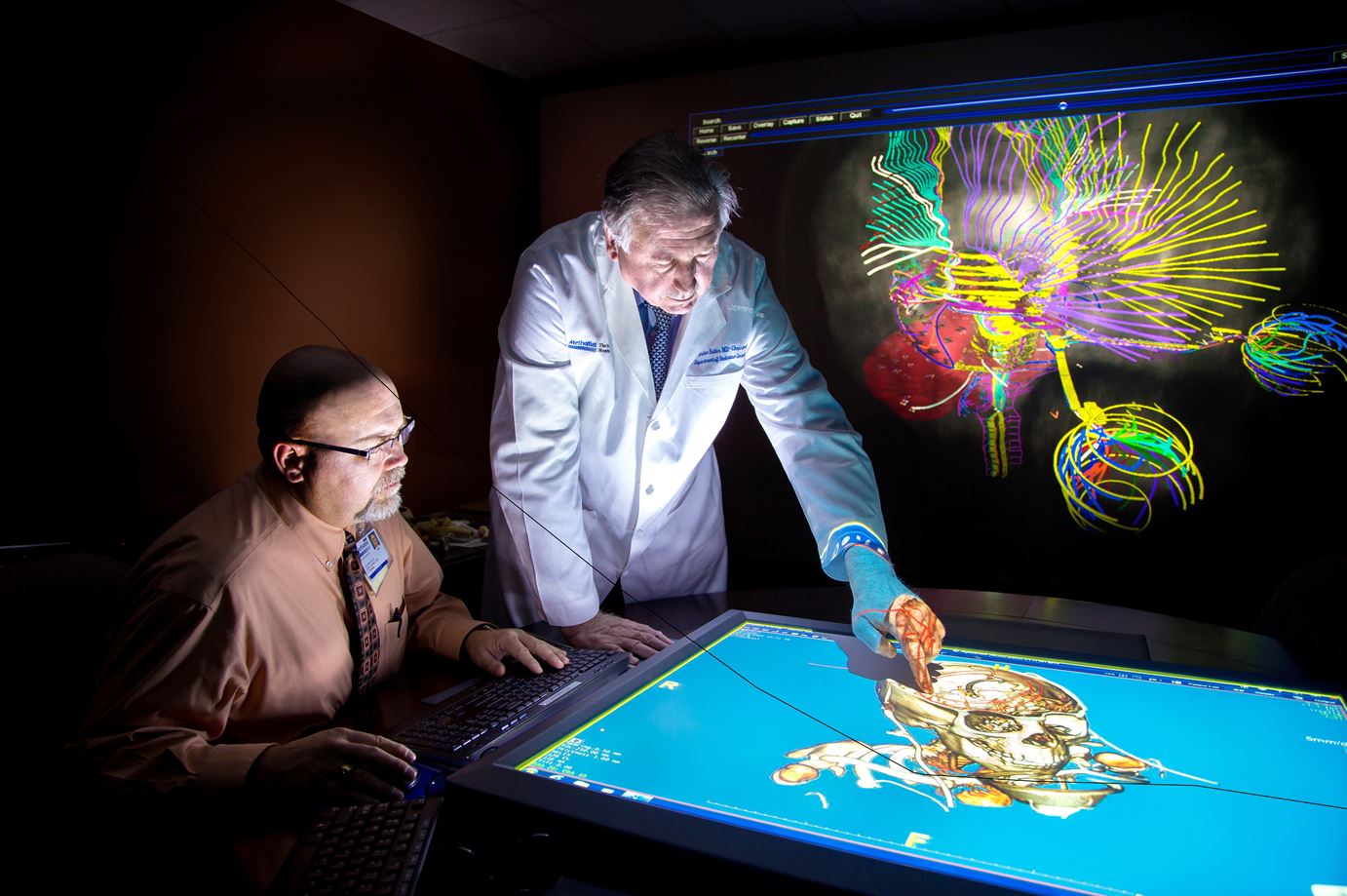
The technology is capable of producing 3-D, 4-D, 5-D and 6-D functional images, meaning doctors have the ability to see a beating heart in exquisite detail and follow a vein or artery through the body. Physicians can navigate through skin, muscle, bones and organs in order to make precise measurements prior to surgery and assess the results of therapy. It is a very impressive advance and this specific virtual environment is only available at Houston Methodist.
Multidisciplinary collaboration within the Department of Radiation Oncology and with other institutions results in scientific research that is translated to the outstanding clinical care we provide. In addition, our standard of care is routinely assessed and updated to ensure that we continue to offer the most effective services to our patients.
Location
6445 Main Street
Outpatient Center, 24th Floor
Houston, TX 77030
713.441.9948